Do You Always Need a Bone Graft for a Dental Implant? The Truth About Jawbone Density
Originally published: July 2026 | Reviewed by Dr. Michael Berglass, DDS

Most patients do not need a bone graft before a single-tooth dental implant. Dr. Michael Berglass, D.D.S., a fellowship-trained implant dentist at West Palm Beach Family Dental, evaluates jawbone density and socket dimensions using three-dimensional CBCT imaging during a free consultation — and the majority of single-tooth cases proceed directly to implant placement without grafting when bone height exceeds 10 mm and ridge width exceeds 5 mm at the planned site.
Bone grafting becomes clinically necessary only when measurable deficiencies fall below specific dimensional thresholds — not as a default add-on to every implant procedure.
Being told you need a bone graft without imaging proof should raise questions — Dr. Berglass shows you the scan and explains exactly why grafting is or is not necessary at West Palm Beach Family Dental.
A dental bone graft is a surgical procedure that adds bone volume — either harvested from the patient’s own body, sourced from a human tissue bank, or fabricated from synthetic, biocompatible materials — to a section of the jaw where natural bone has resorbed to a level below the dimensional threshold required for stable implant placement.
The grafted material acts as a biological scaffold that the body’s osteoblasts (bone-forming cells) gradually infiltrate and replace with native bone tissue through a process the National Institute of Dental and Craniofacial Research’s Bone Grafting clinical summary describes as guided bone regeneration.
Dentists recommend grafting when the existing jawbone cannot provide the surface area and structural density needed to achieve primary stability — the mechanical friction between the implant and the surrounding bone at the time of insertion.
Without adequate primary stability, the implant cannot resist micro-motion during the osseointegration period, and micro-motion exceeding 150 micrometers disrupts bone cell attachment, which determines long-term implant survival.
The clinical question is never whether grafting works — the evidence base confirms its effectiveness. The question is whether the individual patient’s bone architecture actually requires it before implant placement, and that answer comes from imaging, not assumption.
If you’re ready to get started, call us now!
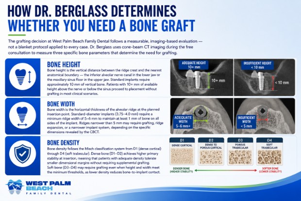
The grafting decision at West Palm Beach Family Dental follows a measurable, imaging-based evaluation — not a blanket protocol applied to every case. Dr. Berglass uses cone-beam CT imaging during the free consultation to measure three specific bone parameters that determine the need for grafting.
Bone height is the vertical distance between the ridge crest and the nearest anatomical boundary — the inferior alveolar nerve canal in the lower jaw or the maxillary sinus floor in the upper jaw. Standard implants require approximately 10 mm of vertical bone.
Patients with 10+ mm of available height above the nerve or below the sinus proceed to placement without grafting in most clinical scenarios.
Bone width is the horizontal thickness of the alveolar ridge at the planned insertion point. Standard-diameter implants (3.75–4.0 mm) require a minimum ridge width of 5–6 mm to maintain at least 1 mm of bone on all sides of the implant. Ridges narrower than 5 mm may require grafting, ridge expansion, or a narrower implant system, depending on the specific dimensions revealed by the CBCT.
Bone density follows the Misch classification system from D1 (dense cortical) through D4 (soft trabecular). Dense bone (D1–D2) achieves higher primary stability at insertion, meaning that patients with adequate density tolerate smaller dimensional margins without requiring supplemental grafting.
Soft bone (D3–D4) may require grafting even when height and width meet the minimum thresholds, as lower density reduces bone-to-implant contact.
| Parameter | Threshold for Direct Placement | Grafting Indicated Below Threshold |
| Bone height | ≥10 mm above nerve / below sinus | Yes — vertical augmentation or short implant alternative |
| Bone width | ≥5–6 mm at insertion site | Yes — lateral augmentation or ridge expansion alternative |
| Bone density | D1–D2 (dense cortical) | D3–D4 may need supplemental support depending on other dimensions |
Dr. Berglass reviews the CBCT model with the patient during the same appointment, pointing to the exact measurements on-screen and explaining why grafting is indicated or unnecessary for that specific jaw anatomy.
Certain clinical presentations require bone grafting before implant placement because the dimensional deficiency is too significant for alternative strategies to overcome safely.
Dr. Berglass identifies these cases during the free consultation and presents grafting as a conditional line item with transparent pricing rather than a default procedure.
Patients who lost teeth more than two to three years ago without receiving implants or prosthetic replacements often present with advanced alveolar resorption.
The American Academy of Periodontology’s Regenerative Procedures position paper states that untreated extraction sites lose 40–60% of ridge width within the first three years after tooth removal — a rate that frequently reduces bone to below the 5 mm minimum required for standard implant diameter.
Long-term denture wearers experience accelerated ridge compression from the prosthetic base pressing against bone that receives no root-like stimulation.
Patients who have worn removable dentures for five or more years commonly present with ridges that are too narrow and too short for any standard implant system without preliminary bone augmentation.
Active or recently treated periodontal disease destroys the alveolar bone in an irregular pattern, creating localized defects around the implant site.
Periodontal bone loss produces vertical crater-shaped defects rather than the uniform horizontal resorption seen in standard post-extraction atrophy, and these craters require targeted grafting to create a level bone platform for implant support.
Traumatic tooth loss — from accidents, sports injuries, or failed root canal retreatment — may fracture the buccal bone plate during extraction, eliminating the labial wall that an implant needs for circumferential bone contact.
The U.S. Food and Drug Administration classifies bone graft materials used in these reconstructive scenarios as Class II medical devices regulated under 21 CFR § 872.3930 for dental use.
If you’re ready to get started, call us now!
The majority of single-tooth implant patients at West Palm Beach Family Dental proceed to placement without bone grafting. Patients most likely to qualify for direct placement share common clinical characteristics that Dr. Berglass identifies during the CBCT evaluation.
Recent extractions within the past 6–12 months typically preserve sufficient ridge volume for immediate or near-immediate implant placement, as significant resorption has not yet occurred. Patients who schedule implant consultations shortly after tooth loss benefit from this timing advantage — the sooner the evaluation happens after extraction, the higher the likelihood of adequate bone for direct placement.
Patients with naturally dense bone (D1–D2 on the Misch classification) maintain ridge dimensions longer after tooth loss than patients with softer bone architecture.
Dense cortical bone resists resorption more effectively and achieves higher primary stability at insertion, often allowing implant placement in sites where softer bone of the same dimensions would require supplemental support.
Anterior tooth replacements — incisors and canines — sit in bone that is typically denser and wider than posterior molar sites, making front-tooth implant cases less likely to require grafting than back-tooth cases, where the bone is thinner and proximity to the sinus or nerve canal adds dimensional constraints.
Three clinical techniques allow Dr. Berglass to place implants in patients with moderate bone deficiency without requiring a separate grafting procedure and its associated 4–6 month healing delay.
Short implant systems measure 6–8 mm in length, compared with the standard 10–16 mm, and are specifically designed for patients with limited bone height above the nerve canal or below the sinus floor.
Research published in the Journal of Oral Implantology confirms that short implants achieve survival rates comparable to those of standard-length implants when proper diameter selection compensates for reduced length and when bone density supports adequate primary stability at insertion.
Tilted implant placement angles the post 15–45 degrees from vertical to engage denser cortical bone at the anterior jaw while bypassing posterior bone deficiency entirely.
The technique is foundational to the All-on-4 protocol, in which two tilted posterior implants and two vertical anterior implants support a full-arch restoration without requiring bone grafting in resorbed posterior regions.
Ridge expansion splits the narrow alveolar ridge laterally to create sufficient width for implant placement without requiring an external graft. Dr. Berglass uses specialized instruments to displace the buccal bone plate outward, widening the ridge from within.
Ridge expansion preserves existing bone, avoids donor-site surgery, and often allows immediate implant placement during the same appointment — eliminating both the grafting cost and the months-long healing delay.
Providers who recommend grafting without first evaluating these alternatives may be following a default protocol rather than a patient-specific plan — Dr. Berglass presents every viable option during the free consultation at West Palm Beach Family Dental.
Patients who receive a grafting recommendation from any provider should ask five questions before consenting to the procedure, since grafting adds $400–$1,500 to the treatment cost and 4–6 months to the overall timeline.
Two-dimensional imaging cannot measure bone width — the dimension most frequently cited as the reason for grafting — so a grafting recommendation without three-dimensional confirmation lacks the diagnostic specificity the procedure demands.
Patients deserve to see the numbers that justify the recommendation rather than accept a general statement that bone is “insufficient.” Measurable thresholds — 10 mm height and 5 mm width — exist for a reason.
Providers who do not offer these alternatives may lack the clinical training or equipment to deliver them — not because the patient’s anatomy precludes them. Dr. Berglass evaluates all three alternative strategies before recommending grafting at any case.
Splitting the case across two offices — an oral surgeon for grafting and a restorative dentist for implant placement — creates referral gaps that add cost and delay treatment. Dr. Berglass handles both procedures under one roof.
West Palm Beach Family Dental’s $2,400 all-inclusive implant price covers the post, abutment, and crown; bone grafting, when genuinely indicated, is quoted as a separate conditional line item with clear clinical justification at the consultation.
The practice is located at 1840 Forest Hill Blvd., Suite 204, West Palm Beach, FL 33406. Call (561) 968-6022 to schedule a free consultation with diagnostic imaging.
A bone graft should solve a measurable problem, not pad a bill — Dr. Berglass proves the necessity on-screen or recommends a path that avoids it at West Palm Beach Family Dental.
Yes — most patients receiving a single-tooth dental implant can avoid bone grafting when CBCT imaging confirms bone height of 10 mm or more and ridge width of 5 mm or more at the planned implant site. Dr. Berglass evaluates these measurements during the free consultation and recommends direct placement, alternative strategies, or grafting based exclusively on the diagnostic imaging findings for each individual patient.
How do I know if I have enough bone for a dental implant?
Dr. Berglass measures bone height, width, and density using three-dimensional CBCT imaging during the free consultation at West Palm Beach Family Dental. Standard implants require approximately 10 mm of bone height and 5–6 mm of ridge width. The CBCT model provides precise measurements to determine whether direct placement, alternative strategies, or grafting is clinically appropriate.
What happens if my bone is too thin for an implant?
Patients with ridge width below 5 mm have several clinical options beyond traditional bone grafting. Ridge expansion widens the existing bone without adding external graft material. Short implant systems and tilted placement techniques bypass the deficient area entirely. Dr. Berglass evaluates which alternative best matches each patient’s specific bone architecture during the consultation.
How much does bone grafting cost before a dental implant?
Bone grafting procedures in South Florida typically range from $400 to $1,500 depending on the graft type, defect size, and material used. West Palm Beach Family Dental quotes grafting as a separate conditional line item when clinically indicated, with transparent pricing presented alongside the practice’s $2,400 all-inclusive implant fee during the free consultation.
How long does bone graft healing take before implant placement?
Bone graft healing typically requires four to six months before the grafted site achieves sufficient density and integration for implant placement. During this period, the graft material serves as a scaffold that the body’s osteoblasts gradually replace with native bone tissue strong enough to support implant insertion and long-term stability.
Can a dentist place an implant and bone graft at the same time?
Minor bone augmentation — such as packing particulate graft material around an implant placed in a slightly undersized socket — can occur simultaneously with implant placement when primary stability is achieved despite the marginal deficiency. Dr. Berglass determines simultaneous eligibility based on the insertion torque measurement recorded during the surgical appointment.
What is the difference between a bone graft and ridge expansion?
Bone grafting involves adding external material to a deficient site and requires 4 to 6 months of healing before implant placement. Ridge expansion splits and displaces the existing bone laterally to create adequate width without adding foreign material, often allowing immediate implant placement during the same appointment with no separate healing period required.
Do all dental offices use CBCT imaging before recommending grafting?
Not all dental offices use three-dimensional CBCT imaging before making bone graft recommendations. Some providers rely on two-dimensional panoramic or periapical X-rays that show bone height but cannot measure bone width — the dimension most commonly cited as justification for grafting procedures, a justification patients question.
Why do some offices recommend bone grafts for every implant patient?
Some offices include bone grafting as a default line item rather than a conditional recommendation based on diagnostic imaging findings. Default grafting protocols add $400–$1,500 to the treatment cost, regardless of whether the patient’s bone dimensions actually require augmentation before implant placement.
Is the bone graft evaluation included in the free consultation?
The free consultation at West Palm Beach Family Dental includes all diagnostic steps needed to determine the necessity of a bone graft — clinical examination, periapical and panoramic X-rays, and CBCT imaging when indicated. Dr. Berglass presents the imaging findings with exact measurements and explains whether grafting, an alternative strategy, or direct placement is appropriate.

About Dr. Berglass
Dr. Michael Berglass, a dedicated dentist with over 25 years of experience, specializes in dental implants, cosmetic, and general dentistry in West Palm Beach, Florida. A proud alumnus of Stony Brook School of Dental Medicine (DDS, 1996), Dr. Berglass further honed his expertise with residencies in general practice and dental implants at North Shore-Long Island Jewish Hospital. His journey in dentistry has taken him from New York to North Carolina, and now to Florida, where he continues to offer top-notch dental care. Known for his wit and intellect, Dr. Berglass makes every patient's visit comfortable. Formerly a member of the American Dental Association and the Florida Dental Association, Dr. Berglass remains committed to staying informed about advancements in dentistry and providing exceptional care to his patients. He looks forward to welcoming you to his practice.
About Dr. Cedenos

Dr. Ivan Cedeno is originally from Cuba where he did his dental studies. Having graduated with a DDS diploma gave Ivan and his family great satisfaction, considering he comes from a family of dedicated dental care professionals. After years of practicing Dentistry in Cuba for a few years, Ivan decided to continue his growth journey as a professional in the United States. Early on, Ivan worked as a laboratory technician, which gave him some unique insights that dental school does not teach. Having graduated from the University of Puerto Rico as a DMD, Ivan is allowed to practice in the USA.


